From Tiny Teeth to 3D Printing!
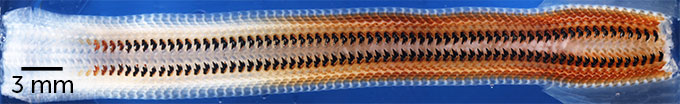
A team of researchers led by Derk Joester (Senior Associate Professor of Materials Science and Engineering at NU), including NUANCE’s very own Paul Smeets, PhD, for the first time discovered a rare mineral hidden inside the teeth of a chiton, a large mollusk found along rocky coastlines. Previously, this strange iron mineral, called santabarbaraite, had only been documented in rocks.
“This mineral has only been observed in geological specimens in very tiny amounts and has never before been seen in a biological context,” said Joester.
To further examine the teeth from the Cryptochiton stelleri, Joester’s team collaborated with a team at Argonne National Laboratory’s Advanced Photon Source, to make use of synchrotron Mössbauer spectroscopy, as well as with Paul Smeets at NUANCE to use transmission electron microscopy, specifically the JEOL ARM300F (S)TEM from our EPIC Facility to capture the below images of the organic matrix of the stylus and the inorganic Santabarbaraite nanoparticles.
In Paul’s words: “Unraveling and understanding the composite structure in the organism set the stage for designing the composite materials with excellent material properties that we manufactured by 3D printing in collaboration with the Hersam Group.”

For more information about this extraordinary discovery, and how the teeth in this mighty mollusk could help unlock further secrets of the biological and mineral world, read the full publication in the Proceedings of the National Academy of Sciences, or the Northwestern Now article!