Focused Ion Beam at EPIC
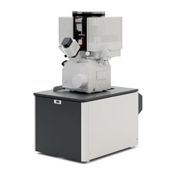
The EPIC-FIB facility will receive a novel well-equipped state-of-the-art focused ion beam – scanning electron microscope (FIB-SEM) in 2025! This instrumentation can combine ultra-high resolution field emission SEM, and FIB material etch and deposition for nanoscale machining, prototyping, 2D and 3D characterization of a wide range of materials ranging from hard to soft matter.
 This Ga+ dual-beam system will amongst others be a dedicated workhorse for conventional and advanced (automated) sample preparation for TEM and atom probe tomography (APT) purposes, to complement our Hydra plasma FIB system.
Stay tuned for more information!
In addition to our state-of-the-art equipment, experienced staff members are on-hand to train and assist users with FIB-SEM work and sample preparation techniques.
EPIC - FIB Highlight
Plasma FIB (PFIB) Tomography Revealing Cellular Ultrastructure
Our Hydra Plasma FIB (PFIB), which acquisition has been graciously supported by the Office for Research at NU, is designed to deliver four different ions as the primary beam, including xenon, nitrogen, oxygen, and argon, each with their own unique applications.
Oxygen PFIB ion milling has been demonstrated to be effective and versatile with biological samples characterization or sample preparation protocols in biological tissues. Here, we show a PFIB Tomography reconstruction of a resin embedded, stained bone marrow-derived cell from a mouse. With a pixel size of several nm and a slice thickness of 20 nm, the ultrastructure of the cell can be readily revealed.
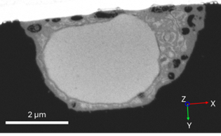 |
| High-resolution backscatter electron (BSE) image of a 2D cross-section within the bone marrow-derived cell. |
| 3D tomographic reconstruction performed within Avizo software of the entire cell, showing segmented nucleus (brown) and vesicles (green). |