"Multi-Scale Materials Science: From 3D to 4D with X-Ray Microscopy"
Sponsored by the NUANCE Center and ZEISS
Jeff Gelb
Senior Applications Engineer, Materials Science
Carl Zeiss X-ray Microscopy, Inc., Pleasanton, CA USA
Friday, August 19
12:00-1:00pm
Northwestern University
Technological Institute
2145 Sheridan Road
Room M164
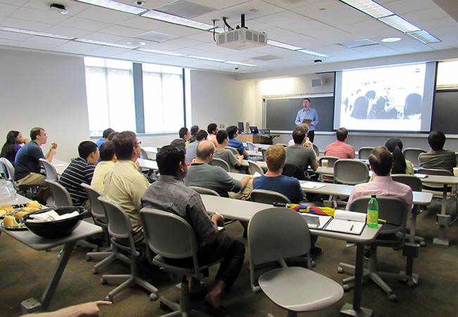
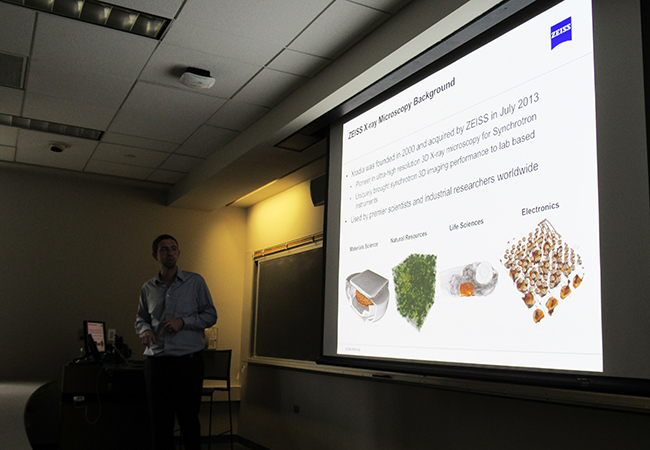
Lunch and learn seminar by ZEISS. Technical discussion of X-ray Microscopy and Correlative Microscopy associated with materials and life science research. Participants learned how this technology can provide nanometer resolution images of a wide variety of samples. Questions were answered about the correlation of XRM data with confocal and other advanced imaging technologies currently available from ZEISS.

ABSTRACT:
X-ray microscopes (XRMs) incorporate a number of X-ray optical elements that have driven resolution and contrast to levels previously unachievable by conventional computed tomography (CT) instrumentation. Analogous to CT, XRM images a specimen without physical sectioning and a complete 3D view of the object is generated. Specifically, XRM adapts advanced detectors that have been developed at synchrotron facilities over the past decades, coupled with high-energy laboratory X-ray sources. The basic system architecture will be reviewed, as well as imaging capabilities such as tunable propagation phase contrast to visualize low-Z materials and diffraction contrast tomography to evaluate crystalline samples. This talk will highlight the imaging capability of XRM with application examples from a variety of fields, including advantages to uniquely characterize the microstructure of materials in situ, as well as the evolution of properties over time (4D). Some recent examples of using XRM in a correlative environment will also be presented, demonstrating how XRM fits into a larger workflow incorporating other characterization instruments, such as SEM, FIB-SEM, and microprobe analysis.

