2016 Image Contest Winners
On October 5, 2016, the finalists of the NUANCE Center Image Contest displayed their stunning creations. Out of 36 submissions using nanoscale imaging instruments available at NUANCE's EPIC and SPID facilities, three winners were elected:
First Place
Dr. Kai He
TEM Facility Manager - Research Assistant Professor - NUANCE Center & Department of Materials Science and Engineering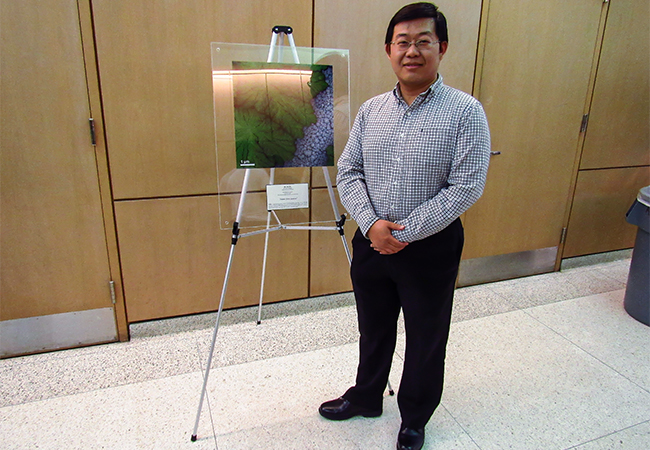
“Nano Fern Leaves”
Dr. He employed a technique called scanning transmission electron microscopy (STEM). He took this STEM image from an electrode material of lithium-ion battery. This reveals the nanoscale dendritic growth of lithium salts inside the electrolyte, which generate the fractal nanostructure similar to a branch of natural fern.
Instrument used to produce image: JEOL 2100F TEM – NUANCE Center/EPIC Facility.
Second Place
Eileen Seo
Graduate Student - Northwestern University - Department of Chemistry - Mirkin research group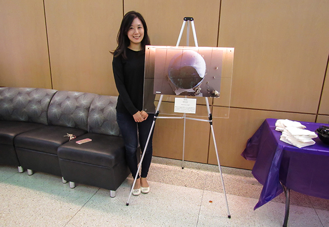
“Discovery of Life”
Eileen created a false-colored SEM image of a cracked glass bead, an impurity found in a DNA-functionalized nanoparticle microcrystal sample, and amorphous silica chunks. Silica encapsulation is a commonly used technique to transfer solution phase DNA-functionalized nanoparticle superlattices to a solid state prior to EM characterization. During the encapsulation process, amorphous silica forms as a side product of nucleation of silane precursors (N-trimethoxysilylpropyl-N, N, N-trimethylammonium chloride and triethoxysilane). The silica chunks (green) in the glass bead are reminiscent of traces of life preserved in a spherical capsule whereas the ones lying outside (brown) are lifeless. Scale bar is 10 um.
Instrument used to produce image: Hitachi SEM 8030 – NUANCE Center/EPIC Facility.
Third Place
Dr. Reiner Bleher
BioCryo Facility Manager - Research Assistant Professor - NUANCE Center & Department of Materials Science and Engineering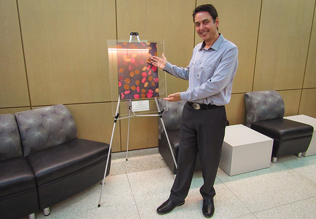
“Osmosis of Red Blood Cells”
Dr. Bleher's image presents Red blood cells at different stages of osmosis after exposure to a hypertonic medium. The micrograph was recorded with the cryo-SEM S4800. The image width is 50 µm. Colors are added for presentation purposes.
Instrument used to produce image: Hitachi SEM S4800 – NUANCE Center/EPIC Facility.
Honorable Mention
Amit Behera
Graduate Student - Northwestern University - Department of Materials Science and Engineering - Olson research group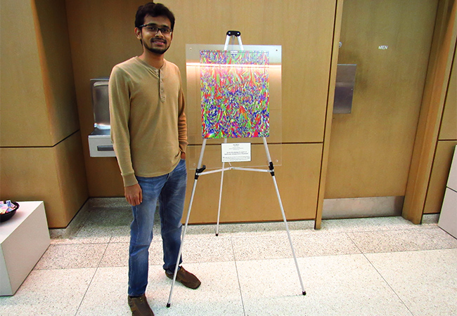
“Grain morphology in layers of Additively manufactured Aluminum”
Amit's image shows the grain morphology across multiple layers of aluminum deposited via additive manufacturing. The build direction is bottom to top for the image. The colors represent crystal orientation according to the color legend at the bottom left corner. Elongated grains are seen across multiple layers due to partial re-melting of previous layers.
Instrument used to produce image: EBSD using the FEI Quanta SEM – NUANCE Center/EPIC Facility.
David Delgado
Graduate Student - Northwestern University - Department of Materials Science and Engineering Shull research group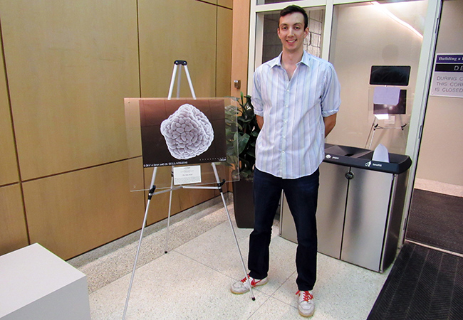
“The Nano Brain”
David: "This image comes from a project that is looking at studying thin film composites made of up a polymer matrix filled with small bits of stuff called carbon nanospheres. Ironically, this image is exactly what I do not want to see: multiple spheres sticking together. To gain a deeper understanding about how these composites behave in their thin film form, these nanospheres need to be spread out and not stuck together. Thus, trying to break up these clusters has been on my brain." Image taken with help from David Collinson.
Instrument used to produce image: Hitachi S4800-II cFEG SEM – NUANCE Center/EPIC Facility.
Jann Grovogui
Graduate Student - Northwestern University - Department of Materials Science and Engineering - Dravid research group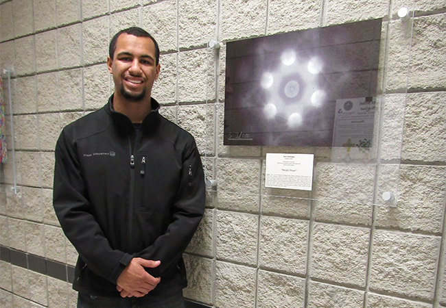
“Purple Phaze”
Jann's image shows a color enhanced Convergent Beam Electron Diffraction (CBED) pattern of a silicon wafer. Diffraction patterns can reveal information about the crystal structure of a material, or reveal the presence of multiple or single phases in a given sample volume. This specific CBED pattern reveals that the silicon is single crystalline and is aligned in the (111) crystallographic direction, which is indicated by the position and spacing of the bright disks. The pattern also reveals a series of lines that can be used to reveal information about atomic spacing and strain in the material.
Instrument used to produce image: Hitachi HT-7700 Biological TEM – NUANCE Center/EPIC Facility.
James Hedrick
Graduate Student - Northwestern University - Department of Chemical and Biological Engineering - Mirkin Research Group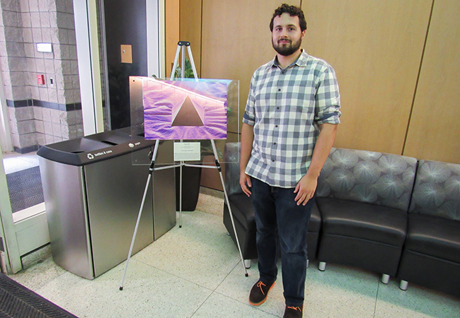
“Lone Glass Pyramid”
James: "Here is an image of the edge pyramidal tip array designed for soft lithography. These pyramids are originally made with a polymeric elastomer, which glass is then deposited onto the surface of the array with PE-CVD. This deposition is done at high temperatures and as it cools down the polymer shrinks. This shrinkage causes buckling to occur, which is visible on the surface around the pyramid. These hard tips will allow for patterning fine densely packed features onto a substrate for a variety of projects. When pressed against the substrate the glass tip will not deform but rather the polymer underneath will decompress creating finer features than a polymeric tip."
Instrument used to produce image: Hitachi SU8030 SEM – NUANCE Center/EPIC Facility.
Dr. Adam E. Jakus
Hartwell Postdoctoral Fellow - Northwestern University - Materials Science and Engineering, Transplant Surgery, Simpson Querrey Institute for BioNanotechnology - Shah research group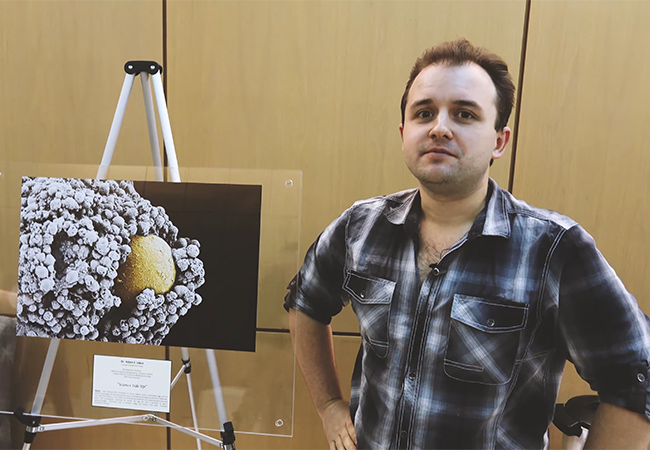
“Science Side Up”
Dr. Jakus' image is of a false Colored SEM micrograph of mouse ovarian follicle (mammalian egg unit) on ovarian tissue paper (A new reproductive system biomaterial developed by the Shah TEAM Lab). This particular follicle is in the middle of shedding the protective and supportive granulosa and theca cells (white/gray), exposing the oocyte, the egg cell that is fertilized by sperm (yellow/orange).
Instrument used to produce image: LEO Gemini 1525 SEM – NUANCE Center/EPIC Facility.
Dr. Yuan Li
Postdoctoral Fellow - Northwestern University - NUANCE Center - Keck-II Facility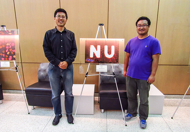
This Raman mapping image is created using SPID LabRam Confocal Raman (NUANCE). The substrate is a “NU” pattern composed of Au@MoS2 core-shell heterostructures and laterally grown few-layer MoS2 flakes. The Raman signal of MoS2 in the “NU” region is significantly enhanced due to the plasmonic effect of Au nanoparticle cores. The inset in the left-bottom corner is a corresponding 3D Raman mapping image.
Instrument used to produce image: LabRam Confocal Raman – NUANCE Center/SPID Facility.
Dr. Magdalena Owczarek
Postdoctoral Fellow - Northwestern University - Department of Chemistry - Stoddart Mechanostereochemistry research group
“Naturally distorted crystal of 4,5-dichloro-2-methylimidazole”
Dr. Owczarek: "An SEM image showing a microstructure of a distorted crystal of 4,5-dichloro-2-methylimidazole. The image allowed us to identify both a bending face of the crystal and, when combined with crystal indexing data obtained from single-crystal X-ray diffraction, structural features that permit the crystal to curve during its formation."
Instrument used to produce image: Hitachi S-3400-II SEM – NUANCE Center/EPIC Facility.
Dr. Xin Wang
Postdoctoral Research Fellow - Northwestern University - NUANCE Center and Department of Materials Science and Engineering - Dravid research group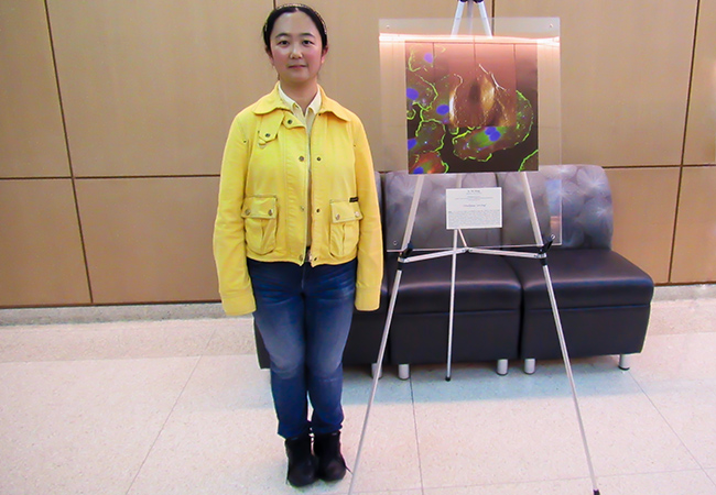
“Chickpeas Serving”
Dr. Wang: "This is correlative image of endothelial cell with AFM PeakForce QNM (Quantitative Nanoscale Mechanical Characterization) and light microscopy image. Green color indicates the immunostaining of focal adhesions, while actin filaments are labeled with red rhodamine-phalloidin, and blue DAPI is used as a counterstain for nucleus identification. AFM PeakForce QNM captures nanomechanical properties—including modulus, adhesion, dissipation, and deformation—while simultaneously 3D topography scanning at atomic resolution. Specific protein distribution, cytoskeleton structure as well as cellular mechanical properties are all integrated in this single image. The cells grown on petri dish substrate look like “color” chickpeas serving as a dish."
Instrument used to produce image: Bruker Resolve – NUANCE Center/SPID Facility.
Congratulations!
More information on the Image Contest, along with detailed images of the eleven finalists, can be found here.
More photos of the event can be found on the NUANCE Facebook Page